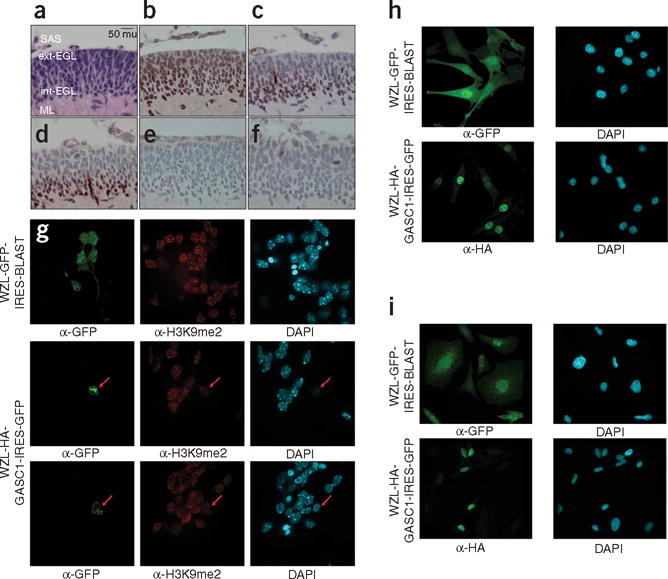
Figure 4.

H3K9 in the developing external granule cell layer. (a) Hematoxylin and eosin (H&E) staining of the P7 murine cerebellum. The external layer of the EGL (ext-EGL), the internal layer of the EGL (int-EGL), the molecular layer (ML), and the subarachnoid space (SAS). Original magnification ×400; scale bar, 50 microns (mu). (b) EHMT1 staining of an adjacent section of the external granule cell layer of the cerebellum. EGL cells are a putative cell of origin in medulloblastoma. (c) Dimethylation of histone 3, lysine 9 (H3K9me2) is seen to be more extensive in the inner, postmitotic layer of the cerebellum, with very little staining in the outer, highly proliferative layer of the EGL. (d) Expression of the cell cycle arrest protein p27Kip1 colocalizes with H3K9me2 in the inner EGL. (e) Monomethylation of H3K9 is not seen by immunohistochemistry in the P7 cerebellum. (f) Rare immunohistochemical staining for H3K9me3 is found in a small subset of mitotic cells of the P7 EGL. (g) Retroviral infection of P7 EGL cells with WZL-GFP shows high efficiency of transduction (infection rate >50%), but only rare cells infected with WZL-HA-JMJD2C could be found (infection rate <1%). EGL cells expressing HA-JMJD2C have decreased levels of H3K9 dimethylation. (h) Viral infection of NIH3T3 cells shows high levels of transduction for both WZL-GFP and WZL-HA-JMJD2C. (i) Viral infection of the medulloblastoma cell line UW228 shows high levels of transduction for both WZL-GFP and WZL-JMJD2C. α-GFP, antibody to GFP DAPI, 4,6-diamidino-2-phenylindole.
