Figure 4.
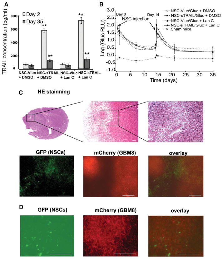
NSCs injected systemically migrate and distribute throughout infiltrating glioblastoma tumors across the blood-brain barrier. Athymic nude mice were stereotactically implanted with 2 × 10⁵ GBM8 cells (~4,000 neurospheres) expressing Fluc and mCherry into the left forebrain. One week later, mice received intravenous injection with 2 × 10⁵ cells of either NSC-sTRAIL/Gluc or NSC-Vluc/Gluc (day 0). NSCs injection was repeated 2 weeks later (day 14). Each group of mice was then divided into two subgroups receiving an i.p. injection (three times per week for 4 weeks) of either 100 μl 1% DMSO or Lan C (1 mg/kg b.wt.). DMSO or Lan C injection was initiated 1 day post-first NSCs injection (day 1). (A): sTRAIL concentration in blood was quantified using ELISA 1 day and 5 weeks after the first injection of NSCs. Data presented as the mean sTRAIL concentration (pg/ml) ± SD (n = 4). **, p <.01 versus control (NSC-Vluc). (B): The fate of injected NSCs was monitored using the Gluc blood assay twice per week. Data presented as the mean value of log (Gluc RLU) ± SD (n = 6). (C): H&E staining showing the invasive foci of glioblastoma cells and fluorescence photomicrograph of GFP (NSC-sTRAIL cells) and mCherry (GBM8 cells) confirms the extensive migration and distribution of NSCs throughout the infiltrating glioblastoma. (D): Higher magnification fluorescence of GFP (NSC-sTRAIL cells) and mCherry (GBM8 cells) confirming the colocalization of NSCs throughout the infiltrating glioblastoma. In (C) and (D), scale bar = 100 μm. Abbreviations: DMSO, dimethyl sulfoxide; GFP, green fluorescent protein; NSC, neural stem cell; TRAIL, tumor necrosis factor-related apoptosis-inducing ligand.

