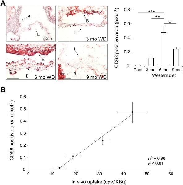
Figure 7.
Inflammation in CAVD. A) Representative examples (left panel) and quantification (right panel) of CD68 expression detected by immunohistochemistry in CAVD. L: leaflet, B: base, mo: months. Scale bar: 100 um. n = 3 in each group, *p < 0.05, **p < 0.01, ***p < 0.001. B) Correlation between CD68 expression and in vivo RP805 uptake in CAVD. R2 = 0.98, p < 0.01.

