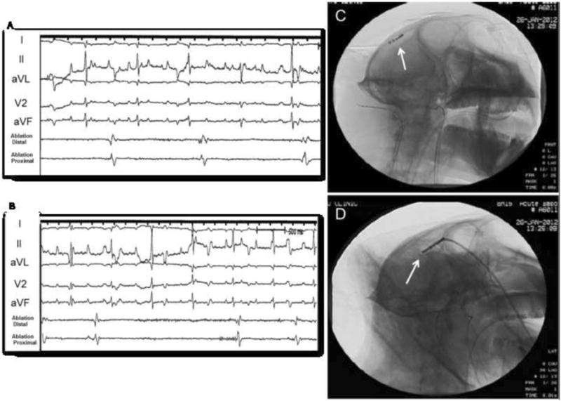
Figure 3. Cerebral activity after Penicillin injection.

Recordings of cerebral activity during partial seizure (A) and after (B) 2500 U of penicillin injection. Panels A and B show cerebral activity presented at the tip of the ablation catheter. Panel C: Cine image from catheter location during seizure activity (AP view); Panel D: Lateral view.
