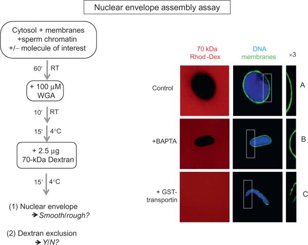
FIGURE 8.2. Nuclear Membrane Assembly and Integrity.
High-speed interphase Xenopus egg extract is mixed with sperm chromatin and membranes and allowed to incubate at room temperature for 60 min. Here, nuclei formed in the presence and absence of a potential inhibitor of membrane fusion are shown. The recombinant protein added to test its role on nuclear membrane assembly and integrity is GST-transportin (as shown in Lau et al., 2009). The integrity of the resulting nuclear envelope was examined with two assays: exclusion of 70 kDa rhodamine dextran (red) and staining with the membrane dye DHCC (green). The latter reveals the presence or absence of a continuous green nuclear envelope. Chromatin is stained with Hoechst DNA stain (blue). For each condition shown, the nuclear envelope is also shown magnified threefold for better viewing (3 × ; right column). (A) The control consists of addition of a protein that does not affect membrane assembly and integrity (here 20 μM GST is used). A smooth nuclear envelope staining is observed and the representative nucleus shown is impermeable to 70 kDa dextran. (B) A similar intact phenotype is observed when 8 mM BAPTA is added to the reaction. (C) Addition of 20 μM GST-transportin prevents vesicle–vesicle fusion, which results in a permeable nuclear envelope. The phenotype is characterized by a rough nuclear envelope staining around the DNA as well as by a permeable nuclear envelope, as determined by the absence of exclusion of the 70 kDa dextran.

