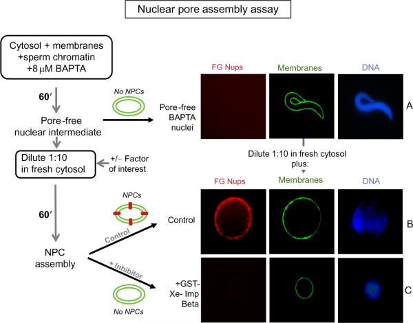
FIGURE 8.3. Nuclear Pore Assembly Assay.
A schematic representation of the assay is depicted at the left. (A) To assemble pore-free nuclear intermediates, sperm chromatin, interphase egg extract, and membranes are incubated in the presence of 8 mM BAPTA for 60 min (Macaulay & Forbes, 1996). The BAPTA intermediate is quite small due to the lack of nuclear pores and resulting lack of nuclear import of vital nuclear proteins. The presence of nuclear pores is detected by staining for FG-nucleoporins (mAb414-TRITC; red), while membranes are detected with DHCC (green), and DNA with Hoechst dye (blue). (B and C) Nuclear pore assembly into these intermediates was tested under different conditions by diluting an aliquot of the pore-free intermediates 1:10 into fresh BAPTA-free cytosol and incubating for a further 60 min. (B) In the control condition, where no inhibitory protein or molecule was added (or when a control protein such as MBP is added), nuclear pore assembly occurred, as seen by the red FG-Nup rim. (C) Addition of 20 μM GST-Xenopus importin beta inhibits nuclear pore assembly, as seen by the lack of FG-Nups at the rim (Harel, Chan, et al., 2003).

