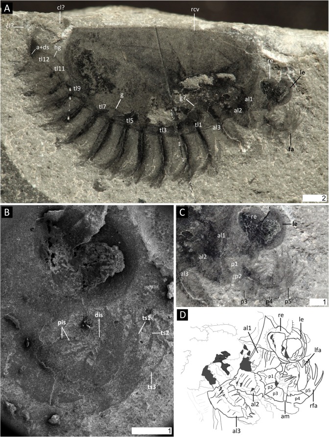
Fig 1. Surusicaris elegans gen. et sp. nov., holotype specimen ROM 62976.
A. Complete view of the part. B. Secondary electron image of anterior area showing details of eyes and frontalmost appendages. C. Close-up of anterior section showing the three pairs of anterior uniramous legs. D. Camera lucida drawing of the “head.” All images were taken under cross-polarized light except in B. Abbr. a+ds: anus+dark stain; alx: anterior limb (1–3); cl?: caudal lobe?; dis: distal inner spine; fa: frontalmost appendage; g(?): gut(?); hg: hindgut; lfa: left frontalmost appendage; le: left eye; px: podomere (1–5); pis: proximal inner spines; rfa: right frontalmost appendage; rcv: right carapacal valve; re: right eye; tlx: trunk limb (1–12); tsx: terminal spine (1–3). Scale numbers in mm.

