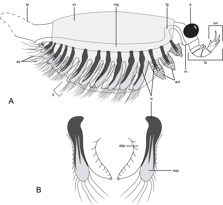
Fig 3. Diagrammatic reconstruction of Surusicaris elegans gen. et sp. nov in profile view.
For clarity, exopods are figured in light grey and caeca in black. A. Habitus. Only the right appendages are drawn and the distalmost segment of the frontalmost appendage is here hypothetically subdivided into three additional segments, based on the anomalocaridid morphology. Exopods are appressed onto the endopod posteriorward to show the tripartite branching of the caeca. The tailpiece is conjectural. B. Antero-posterior view of trunk limbs, with exopod opened up. Abbr. aul: anterior uniramous limbs; cv: carapacal valve; e: eye; ebp: endobasipod; es: exopodial setae; exp: exopod; fa: frontalmost appendage; fg: foregut; ic: invasive caeca; m: mouth; mg: midgut; orc: outer raptorial complex; tl: trunk limb; tp: tailpiece.

