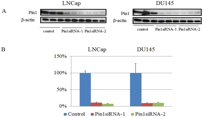
Fig 1.
A. Western blotting using anti-Pin1 antibody and anti- ß-actin antibody as internal controls. LNCap and DU145 cells are transduced with control-siRNA or Pin1 siRNA for 48hr. B. Quantification of bands in Fig 1A. Bars indicate means±S.E. for the ratio of the band intensity of Pin1 to that of ß-actin.

