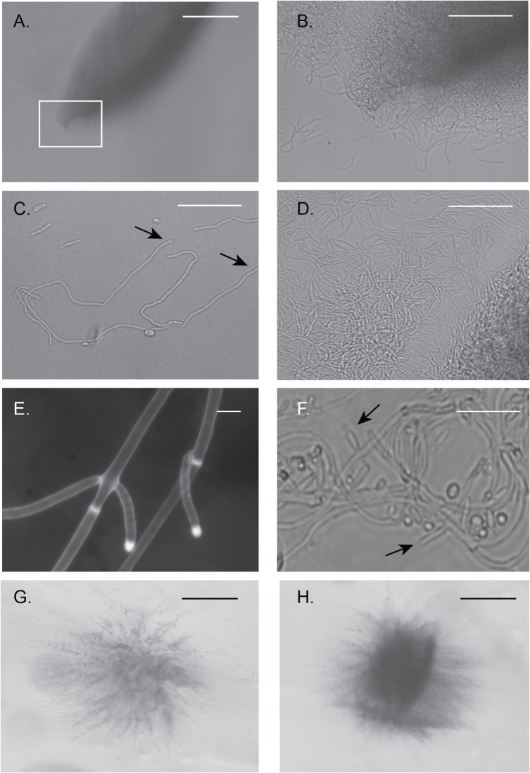
Fig 3. Spindle-shaped colonies reveal fungal tissue formation from embedded HBR1/hbr1 cells.
Hyphal matrix created within a spindle-shaped colony. (A-D). Photomicrographs through a gel slice dissected from a 4 day old embedded culture of CAMPR8 (HBR1/hbr1) and crushed onto a glass slide. (A). A spindle-shaped colony obtained from the deepest part of the agar. Bar = 500 μm (B). Magnification of area boxed in (A) illustrating intertwined hyphae that comprise the spindle colony. Bar = 100 μm (C). Isolated filaments dissected from the gel demonstrating the absence of hyphal branching. The approximate length of the longest filament measured from arrow to arrow is 350μm. Bar = 50 μm. (D). An interwoven mesh of unbranched hyphae taken from a spindle colony. Bar = 50 μm. (E). A filament in the initial stages of branching dissected from a spindle colony and stained with calcofluor white. Note the absence of constrictions at the branch junctions and in the parental filaments, arrows. Bar = 5 μm. (F). Formation of hyphal branches and yeast production from a 10-day old culture. Arrows indicate selected lateral branch points. (*), yeast cell from lateral branch. Bar = 25 μm. (G and H). Initial stages of spindle-shaped colony formation photographed through an agar gel slice. Bar = 500 μm.

