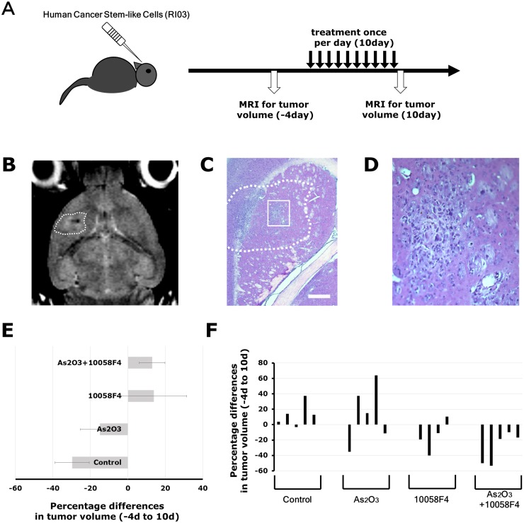
Fig 7. Arsenic trioxide and 10058F4 combination treatment efficiently regressed established gliomas.
Experimental Design. GBM CSCs (RI03) CSCs (5 × 10⁴ cells) were implanted intracranially into SCID mice. Two months after transplantation, tumor growth was monitored by MRI. Four days after tumor size measurement, Arsenic Trioxide (2.5 mg/kg), 10058F4 (25mg/Kg) or both were administered by i.p. injection once a day for 10 days. After 10-day drug treatments, tumor sizes were again measured. Representative images of T2-weighted MRI. The region of interest used to calculate the volume of brain tumor is indicated by a dashed line. (C)-(D) Representative photographs of hematoxylin / eosin staining of intracranial xenograft brain tumors. The boxed area in (C) is magnified in (D). Scale bar = 500μm. (E)-(F) Changes in tumor volume after 10-day treatment with arsenic trioxide and 10058F4 relative to the starting tumor volume for each individual mouse. Each bar represents a volume change of an individual mouse. The data in (E) is shown as the mean ± SD of the data for each individual mouse in (F).

