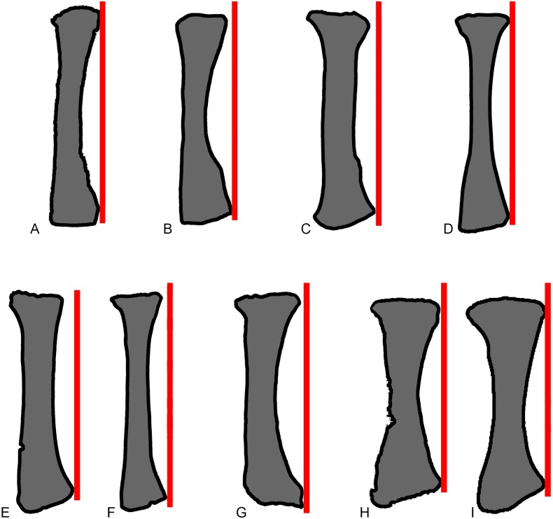
Fig 10. Comparisons of sauropod radii in anterior view.
Exemplar profiles of sauropod left radii in anterior view: A, Mamenchisaurus youngi (ZDM 0083 [107]); B, Ferganasaurus (PIN 3042/1 [101]); C, Apatosaurus louisae (CM 3018 [104]); D, Camarasaurus grandis (YPM 1901 [100]); E, Haestasaurus (NHMUK R1870); F, Giraffatitan (MfN MB.R 2181 [87]); G, Epachthosaurus (UNPSJB-PV 920, based on a photograph by PDM); H, Diamantinasaurus (AAOD 603 [80]); I, Neuquensaurus (MLP-CS 1169 [90]). The red lines are drawn parallel to the vertical long-axis of each radial shaft, at a tangent to the lateral tip of the distal end. F-I are right radii that have been reversed in order to facilitate comparison. Profiles not drawn to the same scale.

