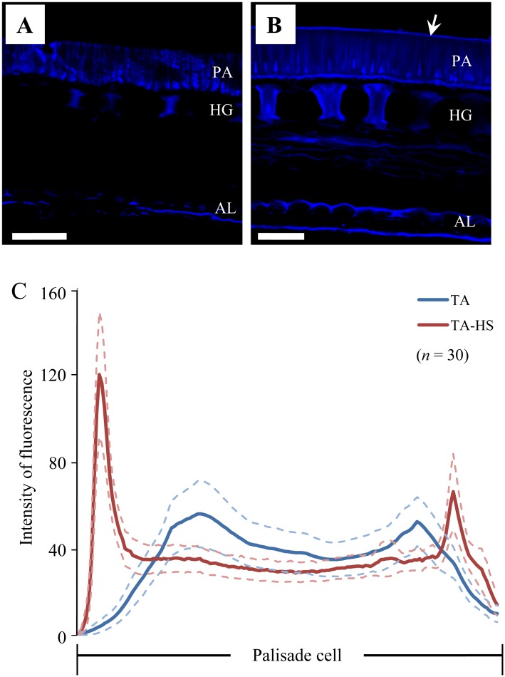
Fig 5. Calcofluor white staining in micro-sections of seed coat.
Cross-sections of seed coats from the dorsal side of seeds of permeable cultivar Tachinagaha (A) and its impermeable NIL (TA-HS) (B) were stained with 0.1% calcofluor white solution and examined under UV illumination by confocal microscopy. Fluorescence indicates accumulation of β-1,4-glucan derivatives in palisade cells (PA), hourglass cells (HG), and outer and inner layers of the aleurone layer (AL). A fluorescent line (arrow) is observed in the outer layer of palisade cells in TA-HS (B), but not in Tachinagaha (A). C, Intensity of fluorescence averaged over 30 positions per genotype at the same relative positions along the long axis of the palisade cell. Blue and red lines are averages for TA-HS and Tachinagaha, respectively, with the maximum errors of estimates at 95% (light blue or pink dashed lines). Scale bars in A and B = 50 μm.

