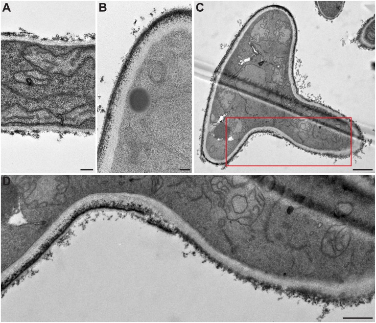
Fig 3. Cell wall modifications during germination of S. boydii under transmission electron microscopy.
(A) hyphal cell wall; (B) conidial cell wall; and (C) cell wall of a germinating conidium. (D) Enlarged part of (C) highlighting the cell wall structural modifications during germination, particularly the electron dense outer layer being less continuous at the surface of the hyphal part of germ tubes. Bars: 0.2 μm in A and B; 1 μm in C; and 0.5 μm in D.

