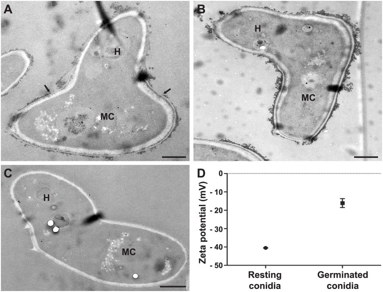
Fig 4. Surface charge modifications during germination of S. boydii by ferritin labeling and zeta potential measurements.
TEM images of germ tubes labeled with cationized ferritin (A), germ tubes treated with neuraminidase prior cationized ferritin labeling (B) or germ tubes incubated with native ferritin (C). (D) Comparison of the surface electrostatic charge of resting and germinated conidia calculated from the electrophoretic mobility of 10 000 cells using Zetasizer Nano ZS (P = 0.0005). H: hyphal part of germ tube; MC: mother cell of germ tube. Bars: 1 μm.

