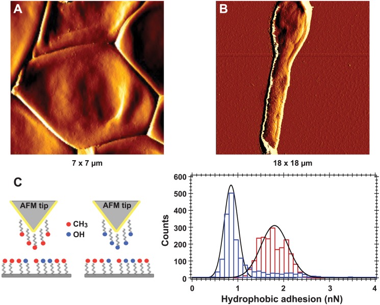
Fig 7. High resolution imaging and chemical force spectroscopy analysis of S. boydii resting conidia and germ tubes.
AFM amplitude images of a resting (A) or germinated (B) S. boydii conidium. (C) Left, scheme for chemical functionalization of AFM tips. Gold-coated tips were modified with CH3-terminated alkanethiols or OH-terminated alkanethiols. (C) Right, histograms of hydrophobic adhesion forces measured on the surface of a resting conidium (1.8 ± 0.3 nN, in red) and the hyphal part of a germinated conidium (0.85 ± 0.15 nN, in blue).

