
Fig. 1.
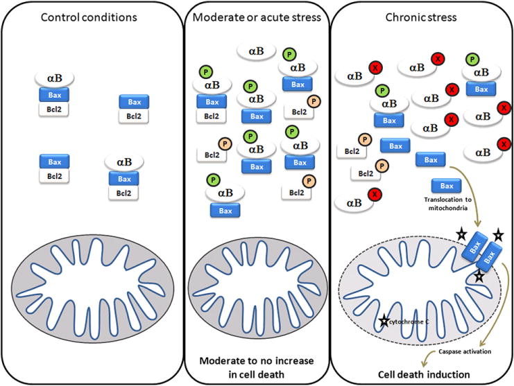
Model of alpha-crystallin functions during chronic (protective) and acute (impaired) stress conditions. In control conditions, anti-apoptotic proteins such as bcl-2 interact and prevent the activation of pro-apoptotic proteins such as Bax. When cells are exposed to a moderate or acute stress, different signaling pathways can be activated leading to the inactivation of the anti-apoptotic member of that complex. Parallel pathways lead to increase AlphaB-crystallin (and alphaA-crystallin) expression as well as anti-apoptotic activity (including phosphorylation) through interaction and inhibition of pro-apoptotic proteins such as Bax and Bcl-Xs. In chronic stress conditions such as diabetes, retinopathy of prematurity or uveitis, alpha-crystallins undergo post-translational modifications such as nitration, glycation, deamidation which inhibit their anti-apoptotic function and lead to cell death induction. (P: phosphorylation; X: unidentified post-translational modification).