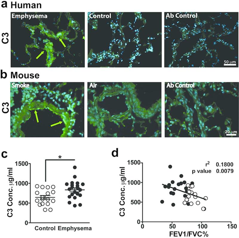
Figure 6. Detection of complement 3 in human and mouse emphysematous lungs.
(a) Representative fresh frozen lung tissue obtained from emphysema or control subjects were stained with anti-C3 or non-immune control antibodies (far right panel). Green fluorescence was achieved by Alexa Fluor 488 (AF488) conjugated secondary antibody and blue fluorescence indicated 4',6-diamidino-2-phenylindole (DAPI) nucleus staining. Figures show overlay of green and blue fluorescence. Scale bar: 50 m. Green arrows indicate C3 deposition. (b) Representative paraffin embedded lung tissue from mice exposed to 6 months of cigarette smoke or air that were stained with anti-C3 or non-immune control antibodies (far right panel). Green fluorescence was achieved with AF488 conjugated secondary antibody and blue fluorescence resulted from DAPI nucleus staining. Figure show overlay of green and blue fluorescence. Scale bar: 20 m. Green arrows indicate C3 deposition. (c) Plasma from control (n=18) and emphysema patients (n=22) were used in an ELISA to measure C3 concentration. **P<0.01, as determined by the Student t test. (d) Correlation of plasma C3 concentration compared with FEV1/FVC%. Solid dots represent individual patients with emphysema and open dots represent individual control patients. P value and r2 were obtained by linear regression model. Results are represented as mean±s.e.m.

