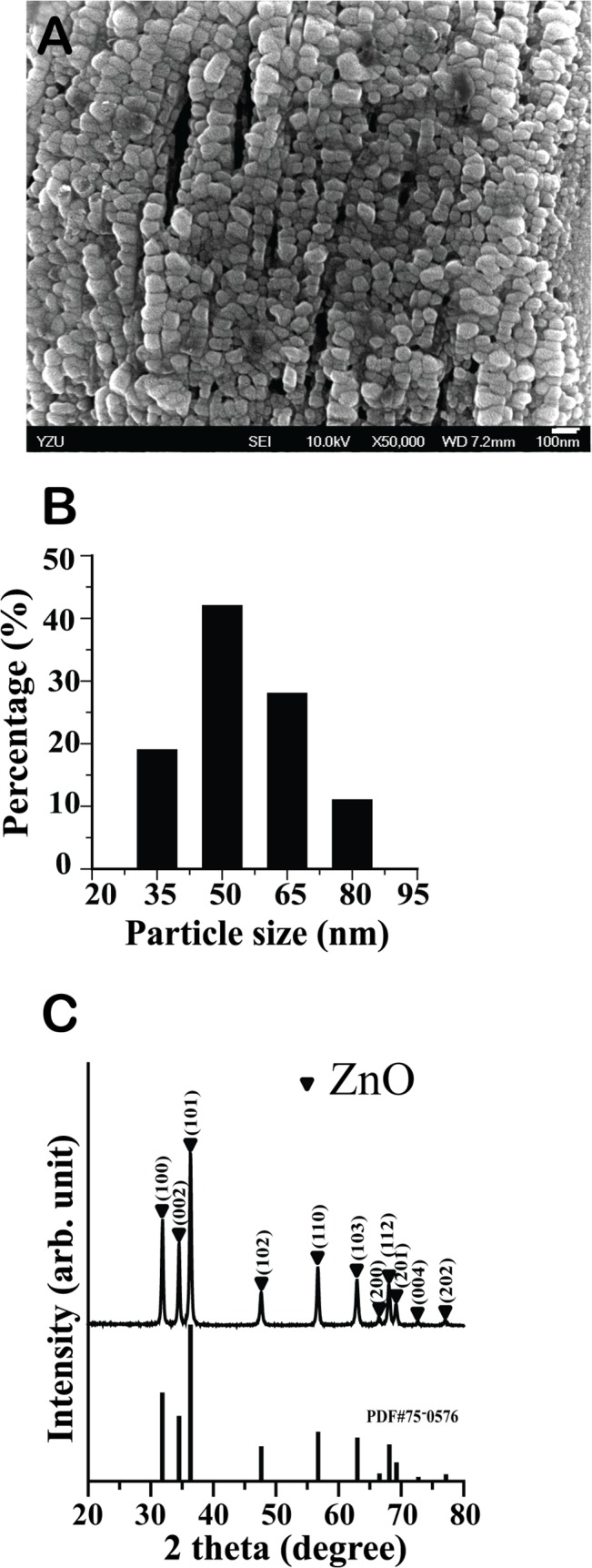
Fig 1. Synthesis and morphological analysis of ZnO NPs.

A. Scanning electron microscope image of ZnO NPs used in this study; white bar: 100 nm. B. Size distribution of ZnO NPs. C. X-ray diffraction patterns of ZnO NPs synthesized by the sol-gel method.
