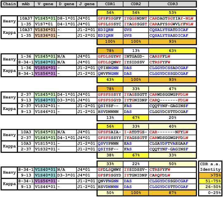
Fig 7. Comparison of CDR regions of the anti-V3 loop mAbs.
The heavy and light chains of the seven V3 loop-positive mAbs were aligned for analysis. Comparison was done based on peptide reactivity shown in Fig 6. Percentages indicate % amino acid identity between the two CDR being compared.

