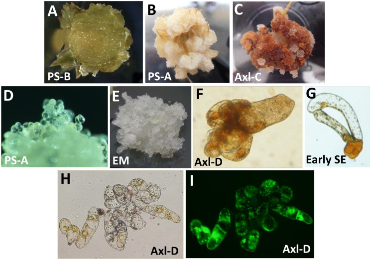
Fig 2. Morphology of tissues induced from primordial and axillary-shoot explants of radiata pine.
A) PS-B explant cultured on EDM Pic for 6 weeks (5x). B) PS-A apical shoot explant cultured on EDM-NAA for 6 weeks (4x). C) Axl-C axillary shoot explant cultured on EDM-Pic for 9 weeks (4x). D) PS-A callus composed of loosely associated round cells growing on EDM-4 (100x). E) EM induced from immature zygotic embryos (5x). F) Axl-D cell aggregate stained with KI (400x). G) EM-A early stage somatic embryo stained with KI (400x). H) Bright-field and I) fluorescence microphotographs of an Axl-D cell aggregate stained with fluorescin diacetate (200x).

