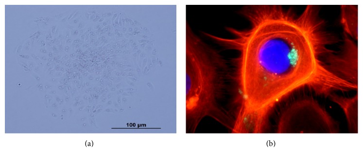
Figure 1.

Normal human thyroid follicular cells in primary culture. (a) Thyrocytes in monolayers showing characteristic morphology by phase contrast microscopy and (b) immunocytochemistry of actin with phalloidin-fluorescein isothiocyanate (red), nuclei stained with DAPI (blue), and thyroperoxidase protein (green).
