Figure 1.
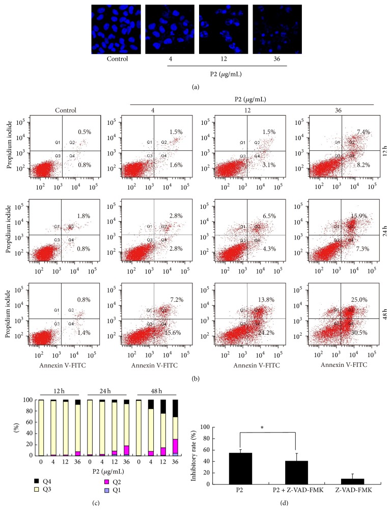
P2 effectively induced apoptosis of HeLa cells. (a) Morphological changes in HeLa cells after treatment with P2 (4, 12, and 36 μg/mL) for 48 h. Cells were stained with DAPI and pictures were taken by LSCM. (b) Apoptosis analysis by FCM for annexin V/PI staining. HeLa cells were treated with P2 at different concentrations (0, 4, 12, and 36 μg/mL) for 12, 24, and 48 h. (c) Histogram of apoptosis rates by P2 in the apoptosis-inducing test. (d) Antiproliferation of P2 on HeLa cells in a caspase-dependent manner. Cells were incubated with 20 μM of Z-VAD-FMK for 1 h before treatment with P2 (12 μg/mL) for 48 h. Cell inhibitory rate was assessed by MTT assay. Compared with P2 single treatment, ∗ P < 0.05 (n = 3).

