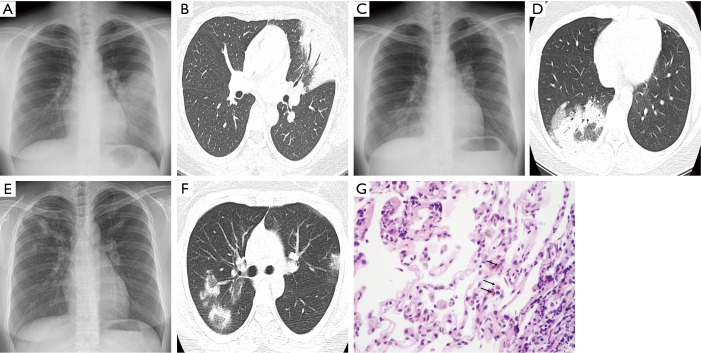
Figure 2.
Chest X-ray (A) and computed tomography (CT) (B) during the first episode of alveolitis (4 months after completion of radiotherapy), showing airspace consolidation in the lingular segment of the left upper lobe (LUL). Chest X-ray (C) and CT (D) during the second episode (7.5 months after radiotherapy) showing patchy airspace infiltrates and consolidation in the lateral and posterior segments of the right lower lobe (RLL) and clearance of the previous LUL lesion. Chest X-ray (E) and CT (F) during the third episode (11 months after radiotherapy) showing multiple airspace consolidations in both upper lobes and clearance of the previous RLL lesion. Transbronchial lung biopsy specimen (G) showing chronic inflammation with several eosinophils (arrows) (H&E stain, ×400).

