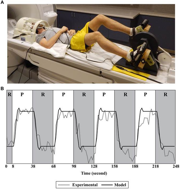
FIGURE 1.
Set-up and fMRI processing. (A) Subject positioned for pedaling during functional magnetic resonance imaging (fMRI). (B) A single run consisted of 30 s of pedaling followed by 30 s of rest, repeated four times. The pedaling portion of the run (P) is shown in white; rest (R) is shown in gray. Recorded data (Experimental) were fit to a canonical function (Model) only during the rest portion of the run. Recorded data (Experimental) represent the time series of a single voxel in the cortex. See Mehta et al. (2009, 2012) for details.

