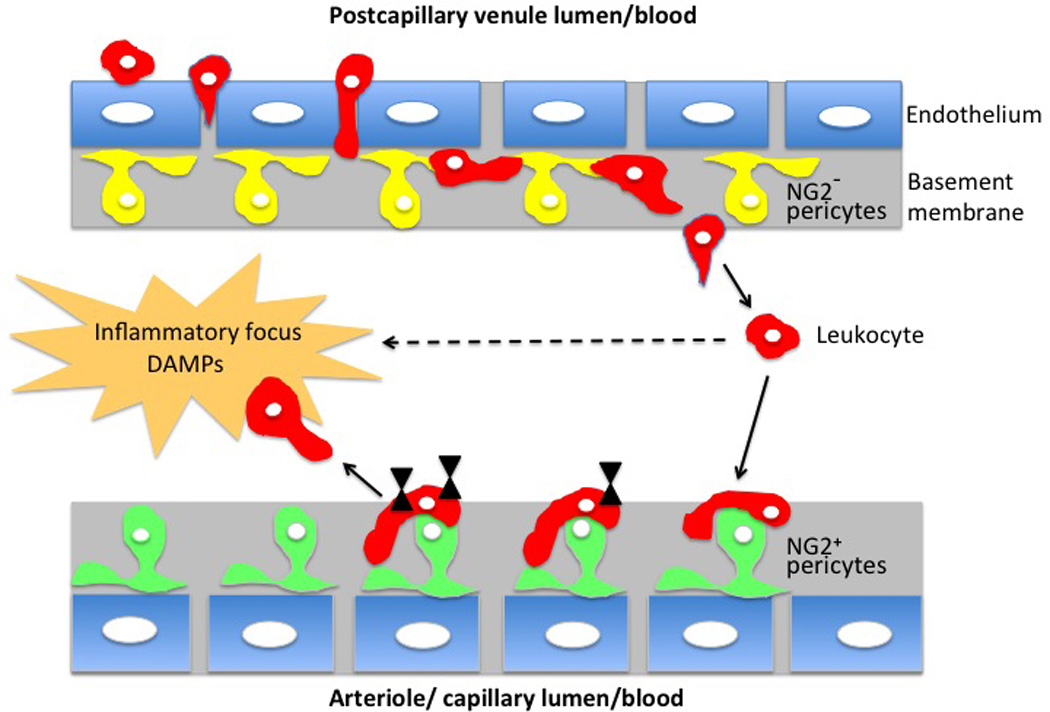
Figure 3. Pericytes participate in endothelial wall breaching by leukocytes, sensitize them and direct them toward the inflammation foci.

Depending on the type of microvessel there are two subtypes of pericytes: venules are covered by pericytes lacking NG2 marker (yellow), and arterioles and capillaries are covered by NG2 positive pericytes (green). During tissue inflammation the leukocytes (red) enter the venular wall and after squeezing between endothelial cells (blue) migrate (abluminal migration) along the processes of NG2− pericytes until they find large inter-pericyte gaps where they are able to exit (extravasate) the vessel wall. Leukocyte migration is ICAM-1, Mac-1 and LFA-1 dependent and a selection of the large-size exit gaps is TNF/IL-1β dependent. Subsequently, after exiting from the venules some of the leukocytes are attracted toward ICAM-1; MIF positive NG2+ pericytes present on neighboring arterioles and capillaries. Contact with NG2+ pericytes and treking along pericyte processes induce leukocytes to produce matrix metalloproteinses, integrins and FPRs (black triangles), progressively acquiring prosurvival signals, upregulating expression of a promigratory receptors and migrating toward the inflammation sites, which are rich in damage-associated molecular pattern (DAMPs) compounds.
