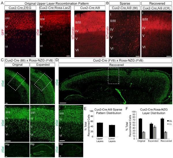
Figure 1. The Cux2 genetic locus exhibits variable activity that depends on genetic background.

(A) Coronal sections from Cux2-Cre mice crossed to Cre reporter lines (all on a congenic C57BL/6J background). Note recombination primarily in upper neocortical layers, with some scattered cells in lower layers. B) Coronal sections from Cux2-Cre mice crossed to the Ai9 reporter showing sparse recombination in the homozygous inbred line (left panel) and the recovered original pattern after outbreeding onto the ICR strain (right panel). (C) Coronal sections from Cux2-Cre;Rosa26-NZG mice showing the original (left) and expanded (right) recombination patterns in the neocortex (top, middle) and hippocampus (bottom). Middle panels: enlarged images of boxed areas in top panels. (D) Sagittal section from a Cux2-Cre;Rosa26-NZG mouse showing the recovered original recombination pattern in the neocortex (top, middle) and hippocampus (bottom) after outbreeding Cux2-Cre onto an FVB/NJ background for 5 generations. Middle panel is enlarged image of boxed area in top panel. (E) Quantification (mean ± SEM) of layer distribution of tdTomato+ cells in sparsely recombined Cux2-Cre;Ai9 mice (502 cells from 3 animals). (F) Quantification (mean ± SEM) of the layer distribution of βGal+ cells in Cux2-Cre;Rosa26-NZG mice exhibiting the different recombination patterns in (C) and (D). Scale bars: (A,B) 100 μm; (C) 200 μm; (D) 500 μm (top) and 200 μm (middle, bottom).
