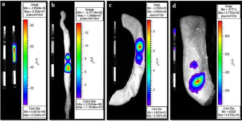
Figure 4.

Representative intra-uterine bioluminescence imaging of labeled spermatozoa. Spermatozoa were labeled or not with 1nM QD-BRET (QD1−), loaded into 0.5-ml plastic straws and imaged inside and outside each reproductive tract sections. Bioluminescence emissions were captured ex vivo, outside (a) and on the surface of oviduct (b), uterine horn (c), and uterine body (d) sections. The ratios of outside over inside luminescence signals (×100) were used to express and interpret data between sections.
