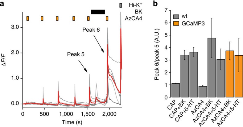
Figure 6. Serotonin and BK sensitized TRPV1 to AzCA4.
Intracellular Ca2+ imaging showed that both BK and 5-HT sensitized TRPV1 to CAP (100 nM) and AzCA4 (200 nM), with ultraviolet irradiation (λ=365 nm, 5 s) in cultured wt DRG neurons. (a) After the application of BK (200 nM), an increased intensity and duration of Ca2+ influx was observed on application of AzCA-4 with ultraviolet irradiation when compared with the previous pulse. The neurons responded to a Hi-K+ solution (100 mM). Shown here are five representative traces (grey) and average ΔF/F value (red). (b) TRPV1 sensitization experiments in wt mouse DRG neurons (grey) and the Trpv1Cre/GCaMP3 mouse line (orange). The results were plotted as the ratio of peak heights for Peak 6/Peak 5 for the wt mouse as follows: CAP with no sensitization agent (n=520); CAP with BK (n=210); CAP with 5-HT (n=175); AzCA4 with no sensitizing agent (n=36); AzCA4 with BK (n=15); and AzCA4 with 5-HT (n=12). For the Trpv1Cre/GCaMP3 mouse line, the results are plotted for the following: AzCA4 with BK (n=28) and AzCA4 with 5-HT (n=10). Error bars were calculated as s.e.m.

