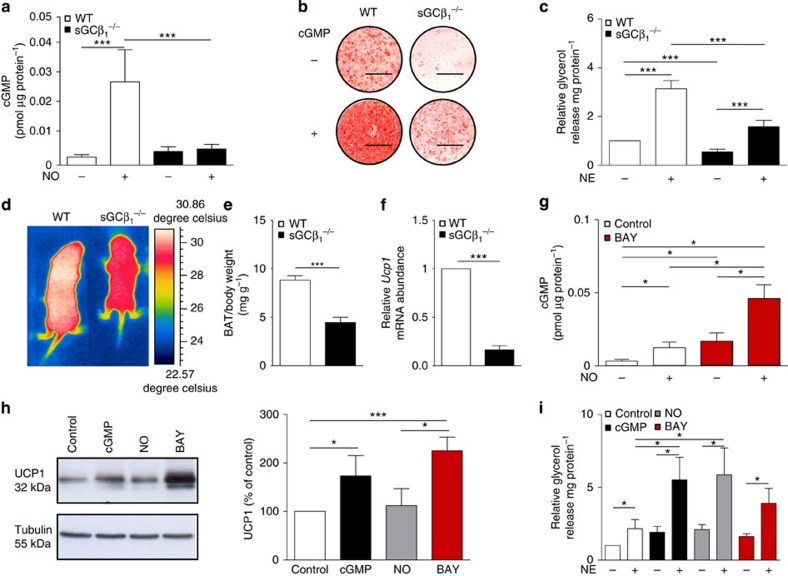
Figure 1. sGC is crucial for development and function of BAT.
(a) Basal and NO-stimulated cGMP content of BA, n=5 independent cell cultures. (b) Oil Red-O stain of WT and sGCβ1−/− BA differentiated in the presence or absence of 200 μM 8-pCPT-cGMP (cGMP), scale bar, 1 cm. (c) Lipolysis in WT and sGCβ1−/− BA under basal and NE-stimulated conditions, n=5–7 independent cell cultures. (d) Representative thermographic image of newborn sGCβ1−/− and WT mice. (e) Weight of BAT from newborn WT and sGCβ1−/− mice, n=8 mice per genotype. (f) Ucp1 gene expression in BAT of newborn mice, n=6 per genotype. (g) Basal NO- and BAY-stimulated intracellular cGMP levels of WT BA, which were incubated for 15 min with the indicated compounds, n=4 independent cell cultures. (h) UCP1 expression of WT BA differentiated in the presence of cGMP, 50 μM DETA/NO (NO) or 3μM BAY, representative western blot (left) and densitometric quantification normalized to loading control tubulin (right), n=3–4 independent cell cultures. (i) Lipolysis in BA under basal and NE-stimulated conditions, n=4–5 independent cell cultures. All data were assessed using Student's t-test and are presented as means±s.e.m. *P<0.05; ***P<0.005.

