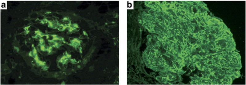
Figure 2.
Immunofluorescence microscopy findings in endocarditis-associated glomerulonephritis. (a) Glomerulus with predominantly mesangial staining by C3 (fluorescein-conjugated anti-human C3; original magnification × 400). (b) Glomerulus with mesangial and capillary wall reaction with C3 (fluorescein-conjugated anti-human C3; original magnification × 400).

