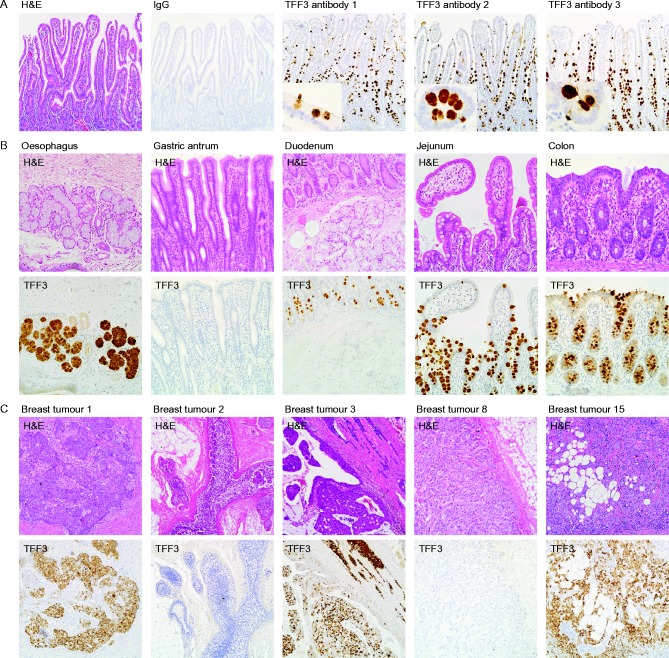
Figure 3.
Immunohistochemical validation of the antibodies against human TFF3 protein. Sections of human ileum (A), oesophagus, gastric antrum, duodenum, jejunum, and colon (B) and of primary breast tumours (C) were stained with haematoxylin and eosin or processed for immunohistochemistry with antibodies against mouse IgG or TFF3 as indicated. The original magnifications for the photomicrographs were ×100 (A and C), ×200 (B) and ×400 for the inserts in (A). A full colour version of this figure is available at http://dx.doi.org/10.1530/ERC-15-0129.

 This work is licensed under a