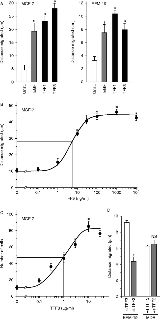
Figure 7.

The effects of TFF3 on breast cancer cell motility and invasion. Monolayers of cells were ‘wounded’ with a pipette tip (A, B and D), and incubated in the absence (unst.) or presence of 10 ng/ml EGF or 5 μg/ml TFF1 or TFF3 for 5 h (A) or the indicated concentration of TFF3 for 7 h (B) or transfected with antisense TFF3 RNA and incubated for 5 h (D). The distance that the wounded edges moved was measured, and the mean distances±s.e.m. of at least four measurements is shown. For the invasion assay, 2.0×10⁴ cells were placed in the upper wells of a microchemotaxis chamber in medium containing 0.01% BSA. Cells were left to migrate on and through 8 μm pore polyvinylpyrrolidome membranes covered with collagen towards the lower wells, which contained the same medium but different concentrations of TFF3. Cells were counted in five high-power fields from triplicate wells, and the means±s.e.m. are shown (C). *Cell movement is statistically significantly higher than for unstimulated cells or lower than for cells transfected with TFF3 siRNA (ANOVA P<0.05).

 This work is licensed under a