Figure 1.
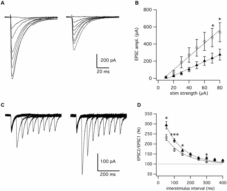
Genetic deletion of Fgf14 suppresses synaptic transmission at parallel fibers to Purkinje cell synapses. (A) Representative superimposed traces of EPSCs evoked by parallel fiber stimulation with 10–80 μA from Fgf14+/+ (left) and Fgf14−/− mice (right). (B) Input-output curve representing EPSC mean amplitude vs. parallel fibers stimulation with linear fitting curves. Note the reduction of EPSCs responses in Fgf14−/− (n = 18, filled triangles) in comparison to Fgf14+/+ (n = 20, open circles). (C) Superimposed traces of EPSCs evoked by paired-pulse stimulation of parallel fibers with interpulse intervals from 50 to 400 ms in Fgf14+/+ (left) and Fgf14−/− mice (right). (D) Time course of paired-pulse facilitation in Fgf14+/+ (n = 14, open circles) and Fgf14−/− mice (n = 18, filled triangles) with exponential fitting curves. Note that PPF at short inter-stimulus intervals, PPF is significantly higher in Fgf14−/− (filled triangles) in comparison to Fgf14+/+ mice (open circles); data are mean ± SEM; *: P < 0.05; ***: P < 0.001.

