
Figure 6.
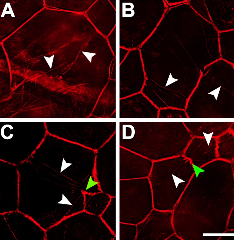
Retinal pigment epithelial cells have stress fibers in AMD-affected eyes. Multiple intracellular stress fibers (white arrowheads) arbitrarily cross RPE cells (A–D), all of which are enlarged. At sites where stress fibers insert, the cytoskeleton appears frayed and thickened ([C, D] green arrowheads). Although RPE cells are found within atrophic areas, only those outside the atrophic area had recognizable actin cytoskeleton and thus stress fibers. Donors: (A, D) 94 years, female, incipient AMD; (B, C) 81 years, male, geographic atrophy. F-actin labeled with AlexaFluor647-Phalloidin. Scale bar: 20 μm.