
Figure 5. Analysis of neuropathology in conditional R1441C LRRK2 transgenic mice.
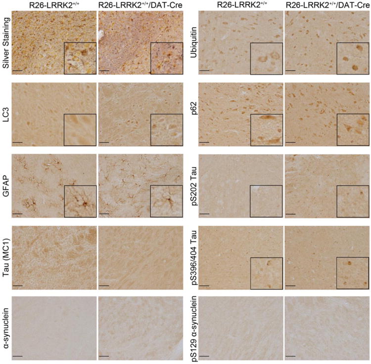
Representative photomicrographs from immunohistochemical analysis of the substantia nigra pars compacta of R26-LRRK2+/+/DAT-Cre and R26-LRRK2+/+ mice at 22 months of age. Sections were processed with Gallyas silver stain or antibodies detecting ubiquitin, the autophagosome marker LC3, the autophagy substrate p62/sequestosome, astrocytic marker GFAP, tau phosphorylated at Ser202 (clone CP13), tau pathological conformation (clone MC1), tau phosphorylated at Ser396/Ser404 (clone PHF-1), total α-synuclein, and α-synuclein phosphorylated at Ser129 (P-α-synuclein). Insets display higher magnification images. Scale bar: 50 μm.