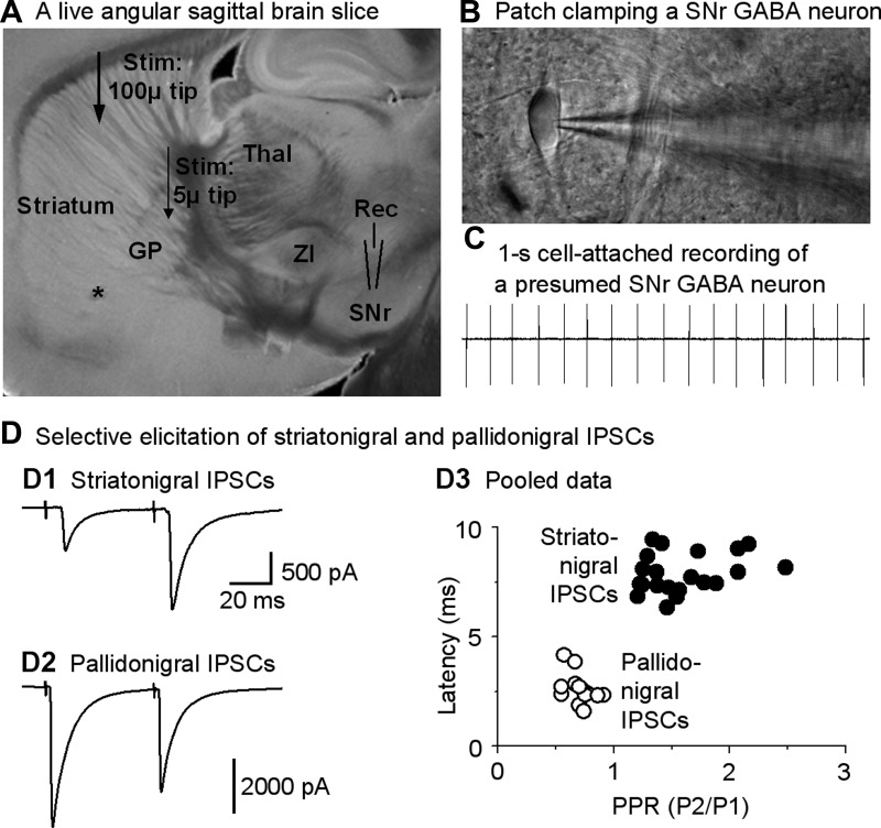
Fig. 2.
Focal stimulation in the striatum or globus pallidus (GP) evokes relatively pure striatonigral and pallidonigral inhibitory postsynaptic currents (IPSCs) in SNr GABA neurons. A: image of a live, 15° angular sagittal brain slice taken with a ×1 objective. The SNr, GP, and striatum are clearly identifiable. Other structures such as the thalamus (Thal) and zona incerta (ZI) are also clearly visible, as marked. B: image taken under a ×60 objective shows a typical SNr GABA neuron being patch-clamped in cell-attached mode. C: typical spontaneous spikes (action potentials, ∼17 Hz) in a presumed SNr GABA neuron recorded in cell-attached mode. D: independent elicitation of striatonigral IPSCs and pallidonigral IPSCs. D1 and D2: example traces of striatum-evoked typical striatonigral facilitating IPSCs and GP-evoked depressing pallidonigral IPSCs, respectively. Timescale applies to both striatonigral IPSCs and pallidonigral IPSCs. D3: pooled data for paired-pulse ratios (PPR; P1 and P2, pulse 1 and pulse 2) and latencies of the first evoked IPSCs induced by a paired-pulse protocol with an interval of 50 ms for striatonigral IPSCs and pallidonigral IPSCs. For striatonigral IPSC data, n = 26 cells from 26 brain slices of 22 mice; for pallidonigral IPSC data, n = 11 cells from 11 brain slices of 11 mice.

