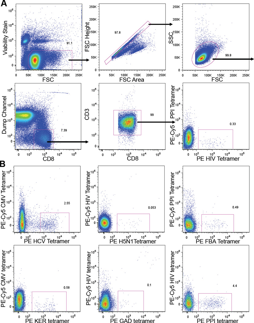
Figure 1. Flow Cytometry of Peptide HLA-A*0201 Tetramer Enriched CD8+ T Cells.
(A) Flow cytometry gating scheme. PBMCs from a HLA-A*0201+ blood donor were concentrated for CD8+ T cells by depletion, followed by HIV:HLA-A*0201 tetramer enrichment over a magnetized column before flow cytometric analysis. Dump channel includes cells labeled with antibodies against CD4, CD14, CD16, CD19, and γδ TCR. In this case, the PE-Cy5 peptide HLA-A*0201 tetramer was only used as control for peptide MHC specific binding.
(B) Representative flow cytometric plots of different peptide HLA-A*0201 tetramer enriched CD8+ T cells. Panels shown are gated on CD8+ T cells. See also Figure S1.

