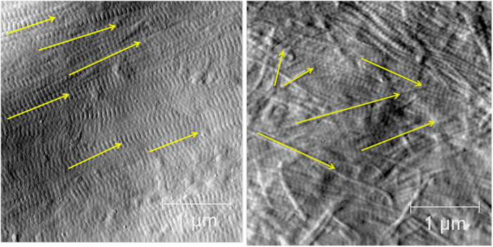
Figure 1.

AFM images illustrating Parallel and Oblique regions of Type I collagen fibrils. (a) Parallel region showing multiple aligned fibrils (yellow arrows); (b) oblique region showing multiple fibrils with varying alignment (yellow arrows). 3.5 × 3.5 μm image.
