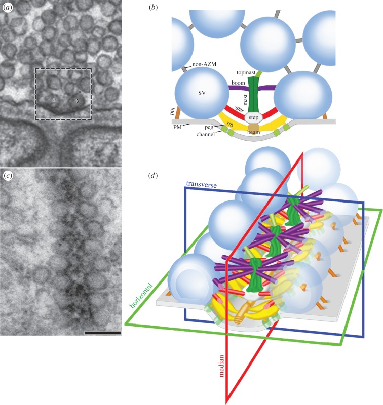
Figure 1.
Arrangement of AZM at active zones on the presynaptic membrane of frog neuromuscular junctions. (a) A 2D electron micrograph from an approximately 80 nm thick tissue section cut in the transverse plane of an active zone. A dense aggregate of macromolecules, constituting the main body of AZM, is seated in the presynaptic membrane's active zone ridge and flanked by synaptic vesicles (asterisks) docked on the presynaptic membrane. The area in the box approximates the area shown in b. (b) Composite diagram of the relationships of AZM macromolecules exposed by electron tomography and viewed in the active zone's transverse plane. Ribs, spars and booms arise from beams, steps and masts in the main body of the AZM to connect to specific domains of the membrane of docked synaptic vesicles (SV) according to their depth/distance from the presynaptic membrane (PM). Pegs link ribs to macromolecules, thought to include Ca2+-channels and Ca2+-activated K+-channels in the presynaptic membrane, while beyond the main body of AZM, the pins link the vesicle membrane directly to the presynaptic membrane. The topmast links the deep end of the mast to an undocked synaptic vesicle. Non-AZM macromolecules connect the membrane of docked synaptic vesicles to the membrane of nearby undocked synaptic vesicles and are similar to filamentous macromolecules that connect undocked synaptic vesicles to each other. (c) A 2D electron micrograph from an approximately 50 nm thick tissue section cut in the horizontal plane of an active zone. The main body of the AZM in this plane is a band. The docked synaptic vesicles are arranged in a single row on each side of it. Scale bar, 100 nm for a and c. (d) A 3D schematic of the active zone showing the same structures, with the same colour code, that are labelled in b, with indicators of the active zone's horizontal, transverse and median planes. (a) and (c) adapted from [20]; (b) and (d) from [21].

