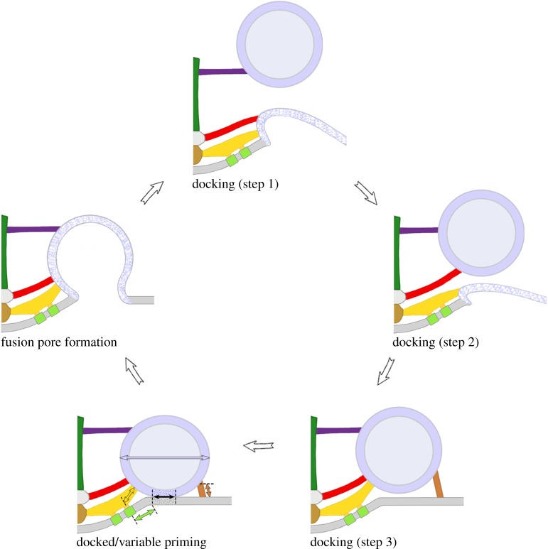
Figure 4.
Model for AZM-mediated docking, priming and Ca2+-triggered vesicle membrane–presynaptic membrane fusion. After a docked synaptic vesicle fuses with and flattens into the presynaptic membrane (blue and white stippled membrane), an undocked synaptic vesicle is directed towards and held in contact with the vacated docking site on the presynaptic membrane by a stepwise progression of stable interactions between it and multiple booms, spars and ribs of the AZM (colour-coded as in figure 1b). Once a synaptic vesicle is docked it undergoes variable priming: the pins and proximal rib segments that link it to the presynaptic membrane shorten and lengthen in dynamic equilibrium (copper and gold double-headed arrows), generating variable force that brings about coordinated variation in (1) the extent of the vesicle membrane–presynaptic membrane contact area and the stability of the lipid bilayers at the contact site (double-headed black arrow), in (2) the proximity of proximal pegs and their associated calcium channels (double-headed green arrow) to the vesicle membrane's Ca2+-sensors and in (3) the synaptic vesicle's eccentricity (blue double-headed arrow) in the plane of the presynaptic membrane. Accordingly, docked synaptic vesicles are most primed when their pins and proximal rib segments are shortest, their vesicle membrane–presynaptic membrane contact areas are largest, their lipid bilayers are most destabilized towards fusion threshold, their associated Ca2+-channels are, on average, in closest proximity to it and they are most eccentric in shape. The membrane of docked synaptic vesicles that are most primed at the moment a nerve impulse arrives has the greatest probability of merging with the presynaptic membrane to form a fusion pore. Fusion occurs while the synaptic vesicle is still attached to the AZM macromolecules. Figure adapted from [20] and [26].

