Figure 2.
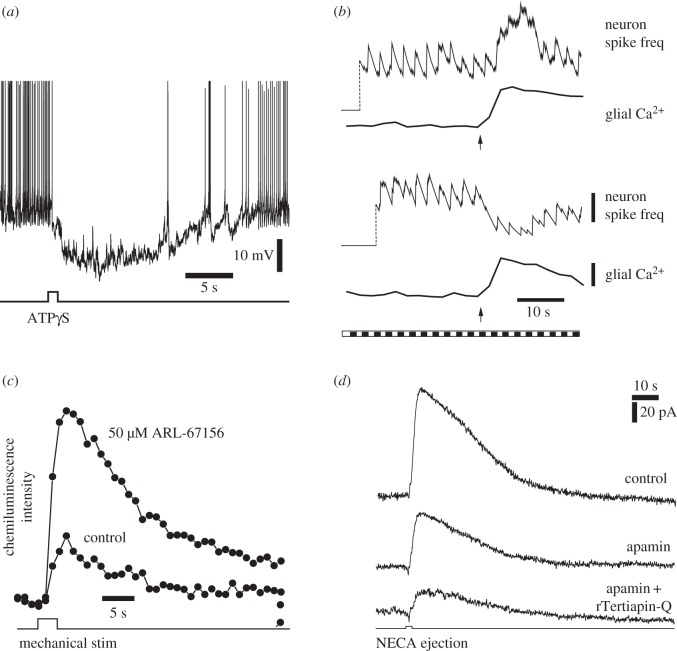
ATP release from glial cells inhibits retinal ganglion cells (RGCs). (a) An intracellular recording from a RGC that displays spontaneous spiking. Stimulation of glial cells by ATPγS ejection results in prolonged hyperpolarization of the RGC and inhibition of spiking. From Newman [9]. (b) Mechanical stimulation of Müller cells (arrows) evokes Ca2+ increases in the glial cells and modulates light-evoked RGC activity. Müller cell stimulation potentiates neuronal activity in some cells (upper traces) and depresses activity in others (bottom traces). The retina was stimulated with a flickering light (bottom trace). Vertical scale bars: running average of neuron spike frequency, 3 spikes per s; glial Ca2+, 20% Δfluorescence/fluorescence. From Newman & Zahs [8]. (c) Mechanical stimulation of Müller cells evokes ATP release into the inner plexiform layer. Extracellular ATP is monitored by the luciferin-luciferase chemiluminescence assay. Addition of ARL-67156, which blocks the conversion of ATP to adenosine in the extracellular space, increases ATP levels. From Newman [9]. (d) Müller cell inhibition of RGCs is mediated by the opening of SK channels and GIRK channels. Ejection of NECA, an A1 adenosine receptor agonist, onto RGCs evokes an outward current in a voltage-clamped RGC. NECA mimics the activation of A1 receptors by ATP/adenosine released from Müller cells. Addition of the SK channel blocker apamin partially reduces the current while addition of apamin and the GIRK channel blocker rTeriapin-Q reduces the current further. From Clark et al. [10].

