Figure 4.
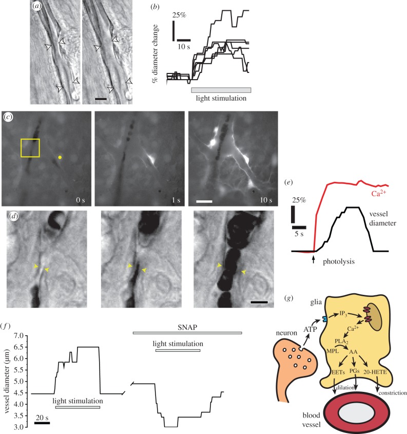
Glial cell release of vasoactive arachidonic acid metabolites mediates neurovascular coupling in the retina. (a) Flicker stimulation dilates arterioles. Photomicrographs of an arteriole in an acutely isolated retina, before and during stimulation. Scale bar, 10 µm. (b) Dilation of retinal arterioles evoked by flicker stimulation. (c and d) Glial stimulation dilates arterioles. (c) Photolysis of caged Ca2+ (yellow dot) evokes a Ca2+ increase in the stimulated astrocyte and in adjacent glial cells. The boxed area is shown in d. Scale bar, 20 µm. (d) Glial cell stimulation results in arteriole dilation. Scale bar, 10 µm. (e) Analysis of the experiment shown in c and d. Photolysis of caged Ca2+ evokes a glial Ca2+ increase and an arteriole dilation. (f) Nitric oxide blocks light-evoked arteriole dilation, revealing a light-evoked constriction. Addition of the NO donor SNAP transforms a light-evoked dilation to a constriction. (g) Summary of glial-mediated neurovascular coupling in the retina. Synaptic release of ATP from neurons stimulates purinergic receptors on glial cells, leading to the production of IP3 and the release of Ca2+ from internal stores. Ca2+ activates phospholipase A2 (PLA2), which converts membrane phospholipids (MPL) to arachidonic acid (AA) which is subsequently metabolized to the vasodilators prostaglandin E2 (PGE2) and epoxyeicosatrienoic acids (EETs), and to the vasoconstrictor 20-hydroxy-eicosatetraenoic acid (20-HETE). (a–f) From Metea & Newman [18] and (g) adapted from Newman [20]. (Online version in colour.)

