Figure 5.
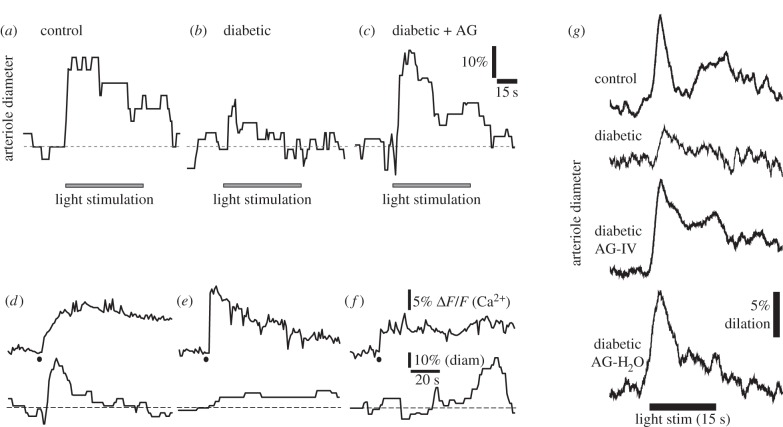
Glial-mediated neurovascular coupling is compromised in diabetic retinopathy. (a) Flicker stimulation dilates an arteriole in an acutely isolated, age-matched control retina. (b) Vasodilations are reduced in the retinas of animals that have been diabetic for seven months. (c) The iNOS inhibitor aminoguanidine (AG) restores flicker-evoked vasodilation in a diabetic retina. (d) Stimulation of glial cells by photolysis of caged Ca2+ (black dot) evokes a glial Ca2+ increase (upper trace) and arteriole dilation (lower trace) in an acutely isolated aged-matched control retina. (e) The glial-evoked vasodilation, but not the glial Ca2+ increase, is reduced in a diabetic retina. (f) Aminoguanidine restores the glial-evoked vasodilation in a diabetic retina. (a–f) From Mishra & Newman [21]. (g) Aminoguanidine reverses the loss of flicker-evoked vasodilation in vivo. Both acute administration of aminoguanidine by IV injection (AG-IV) and chronic aminoguanidine administration through the drinking water (AG-H2O) reverses the loss of flicker-evoked vasodilation in diabetic animals. From Mishra & Newman [22].

