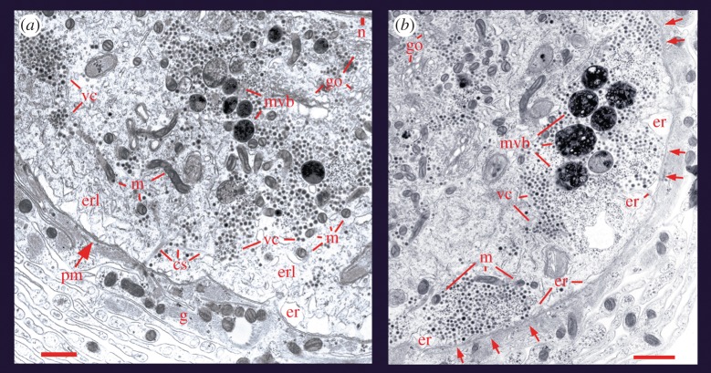
Figure 1.
Ultrastructure of somatic release sites. (a) Electron micrograph of a Retzius neuron in the ganglion that had been fixed after stimulation with 1 Hz trains. The vesicle clusters (vc) remained distant from the plasma membrane (pm), although bound to it through bundles of microtubules (cs). Vesicles appear near mitochondria (m) and endoplasmic reticulum (er). The golgy apparatus (g) and the nucleus (n) are marked on the upper right side. Another population of vesicle clusters and multivesicular bodies (mvb) appear more internally. As explained later in the text, multivesicular bodies are formed after vesicle exocytosis. Retzius neurons are surrounded by layers of a giant glial cell (g). Scale bar, 500 nm. (b) After stimulation with 20 Hz trains, the vesicle clusters appear opposed to the plasma membrane (arrows) and flanked by endoplasmic reticulum and mitochondria. Scale bar, 1 µm. Adapted with permission from De-Miguel et al. [43].

