Figure 2.
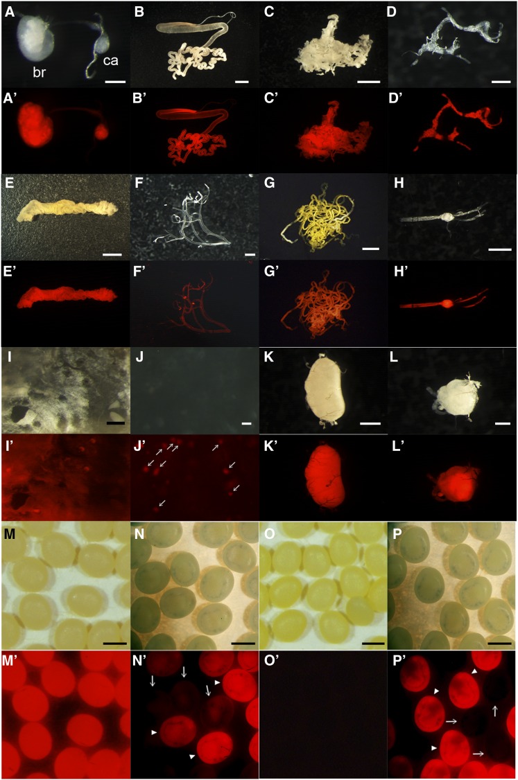
DsRed expression of AyFib-431a in each tissue of final instar larvae (A−L′) or in the embryo (M−P′). (A, A′) Brain and corpora allata. (B, B′) Silk gland. (C, C′) Fat body. (D, D′) Prothoracic gland. (E, E′) Gut. (F, F′) Trachea. (G, G′) Malpighian tubules. (H, H′) Ventral nerves. (I, I′) Epidermis. (J, J′) Hemolymph. (K, K′) Testis. (L, L′) Ovary. (A, B, C, D, E, F, G, H, I, J, K, L) Bright-field images. (A′, B′, C′, D′, E′, F′, G′, H′, I′, J′, K′, L′) DsRed-fluorescent images. br, brain; ca, corpora allata. Arrows in (J′) indicate hemocyte cells. (M, M′, N, N′) Eggs laid by AyFib-431a heterozygous female crossed with w-c male. (M, M′) Just after oviposition. (N, N′) 7 days after oviposition. Arrowheads indicate the putative DsRed+ (AyFib-431a/+) and arrows indicate DsRed− (+/+) individuals. Residual maternal DsRed protein was detected in the putative +/+ embryos. (O, O′, P, P′) Eggs laid by w-c female crossed with AyFib-431a heterozygous male. (O, O′) Just after oviposition. (P, P′) 7 days after oviposition. Arrowheads indicate the putative DsRed+ (AyFib-431a/+) and arrows indicate DsRed− (+/+) individuals. Bars represent 0.25 mm in (A, I), 5 mm in (B, E), 2.5 mm in (C, G), 0.5 mm in (D, L), 1 mm in (F, H, K, M−P), and 0.05 mm in (J).

