
Figure 4-3.
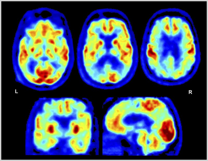
Fluorodeoxyglucose positron emission tomography (FDG-PET) of the patient in Case 4-2. Images displayed in the National Institutes of Health color scale in neurologic orientation. Hypometabolism is noted in bilateral temporoparietal and dorsal prefrontal cortex. Temporoparietal hypometabolism is the FDG pattern associated with Alzheimer disease, while additional involvement of the prefrontal cortex may explain the prominent executive dysfunction experienced by the patient.
L = left; R = right.