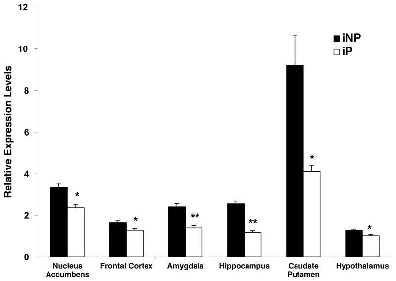
Fig. 1.
NPY mRNA expression is decreased in alcohol-naive P rats compared with NP rats. qRT-PCR analysis was utilized to compare the relative levels of NPY mRNA between alcohol-naive iP and iNP rats in the nucleus accumbens, the frontal cortex, the amygdala, the hippocampus, the caudate putamen, and the hypothalamus. All values were determined using the standard curve method and compared with mRNA expression in the iP hypothalamus, which was arbitrarily designated 1. The graph depicts the mean±S.E.M. of the results from five independent iP and four independent iNP experiments performed in triplicate, using separate preparations of cDNA. Significant differences in regional NPY mRNA expression between iP and iNP rats was determined using the Student’s t-test. * P<0.05; ** P<0.0005.

