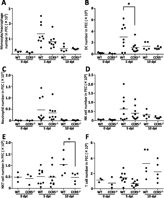
Figure 3.

Migration of peritoneal cells to the site of infection. Peritoneal cells (PEC) were obtained from CCR5−/− mice and C57BL/6 mice (wild-type, WT) at 0, 5 and 10 days post-infection (dpi) with 1 × 10⁶ N. caninum tachyzoites (0 dpi, N = 3; 5 dpi, N = 9 from two independent experiments; 10 dpi, N = 5). The cells were subjected to flow cytometry to determine the absolute number of monocytes/macrophages (A), dendritic cells (B), neutrophils (C), NK cells (D), NKT cells (E) and T cells (F). Cell number per individual (symbols) and mean levels (horizontal lines) are indicated. Data were analyzed by a student’s t-test and compared with values taken on the same day post-infection. *P < 0.05.
