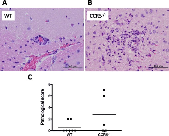
Figure 6.

Pathological analysis of brain tissue. Slight to mild lesions including perivascular cuffs were observed in the C57BL/6 mice (A), and slight to moderate lesions including necrotic focus with glial cell infiltration were observed in the CCR5−/− mice (B). Additionally, the severity of the histopathological lesions was analyzed (C). No significant difference was observed in the pathological score between the two groups (student’s t-test).
